Vascular Lab

Ultrasound Testing

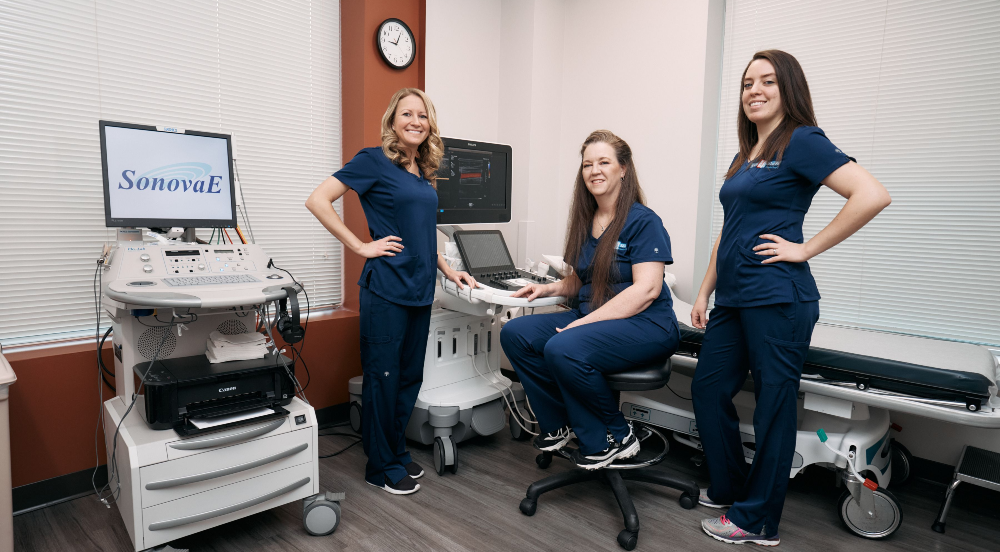
Our state-of-the-art vascular lab has ultrasound imaging equipment that allows us to get precise imaging for an accurate diagnosis and treatment. Ultrasound scans, or sonography, are safe and painless because they use sound waves or echoes, instead of radiation, to produce pictures of the inside of the body.

These pictures allow doctors to target the precise location of the problem area, and can detect conditions occurring in the liver, heart, kidney or abdomen. All image recording happens in real time, so there is no wait for picture development that is required with the use of other imaging procedures.

Tests We Offer
Abdominal Aortic Aneurysm Ultrasound

An abdominal aortic aneurysm occurs when atherosclerosis or plaque buildup causes the walls of the abdominal aorta to become weak and bulge outward like a balloon. The abdominal aorta refers to the part of the aorta—the artery that carries blood from the heart to the legs—between the diaphragm and the legs. Therefore, when a bulge occurs in the abdominal aorta, it is called an abdominal aortic aneurysm.

Ultrasound imaging allows us to assess the condition of the aorta via high-frequency sound waves (ultrasound) that provide detailed information about the blood flow and quality of muscles surrounding this vital tissue.

Carotid Duplex Ultrasound

A carotid Doppler ultrasound is a diagnostic procedure that uses high-frequency sound waves (ultrasound) to generate images of the carotid arteries in the neck. The carotid arteries supply oxygen-rich blood to the brain, which is essential for many neurological conditions. This test allows us to detect any narrowing of the arteries, and also determines how quickly blood flows through them to evaluate a patient’s risk of stroke or other heart conditions.

The carotid Doppler ultrasound is most commonly performed on patients who recently had a stroke, have an abnormal sound in the carotid artery, may have blood clots in the carotid artery, have damage in the walls of the carotid artery or recently had carotid artery surgery.

A carotid Doppler ultrasound usually takes less than 30 minutes to perform.

Deep Vein Thrombosis Ultrasound

Deep vein thrombosis (DVT) is a serious condition that occurs when a blood clot, also known as a thrombus, forms in a vein deep within the body. Clots most frequently form in the legs as a result of several different factors that can affect blood circulation and may involve damage to the inner lining of the vein, slow blood flow or thicker blood.

Deep vein thrombosis (DVT) often goes unnoticed because its symptoms are not always visible. Some people may experience pain, redness and swelling in the legs, ankles and feet. Symptoms may not arise until a pulmonary embolism is present, which can cause chest pain, shortness of breath and dizziness.

Our skilled physicians will perform a physical evaluation, review your medical and family histories, and conduct a series of tests to diagnose your condition. A venous study, using ultrasound technology to produce visible images of the clot, may also be prescribed. If a pulmonary embolism is suspected, additional testing may be performed.

Mesenteric Ultrasound

A mesenteric ultrasound is a non-invasive imaging procedure that uses high-frequency sound waves (ultrasound) to produce images of your mesenteric arteries, which supply oxygenated blood to the intestines, digestive organs and abdominal region. This test is frequently performed on patients who experience abdominal pain or cramping after eating a meal, and typically takes about one hour to perform.

Peripheral Arterial Ultrasound

Peripheral artery disease (PAD), also known as peripheral vascular disease (PVD), occurs when peripheral blood vessels are blocked, hardened and narrowed with plaque, resulting in a condition called atherosclerosis.

An ultrasound using arterial Doppler imaging uses high-frequency sound waves to diagnose peripheral arterial disease (PAD). A Doppler test helps estimate the blood flow through your blood vessels by bouncing high-frequency sound waves (ultrasound) off circulating red blood cells. A regular ultrasound uses sound waves to produce images, but can't show blood flow.

This procedure usually takes about 45 minutes to perform, and patients can return to their regular activities immediately following it.

Renal (kidney) Ultrasound

The renal arteries carry blood to the kidneys, but if these arteries become narrowed or blocked, kidney failure or high blood pressure may occur. A renal artery ultrasound uses high-frequency sound waves (ultrasound) to locate clots or narrowed areas within the arteries. Ultrasound imaging also allows us to determine the size of the kidney and observe the condition of the renal artery, which is determined by measuring the speed of the blood flowing through it. It can also be used to evaluate the severity of a renal artery condition that has already been diagnosed.

Since there are often no symptoms present in the early stages of renal artery disease, this condition may be difficult to diagnose without an effective ultrasound exam.

Ultrasound Tech Team-resz

Locations

Rosedale Center

5233 King Ave, Suite 208 </br / / / / / / / / / / / / / / / / / / / / / />Baltimore, MD 21237

Bel Air Office

BelAirPhoto2023

Images © Google Street View

Search